
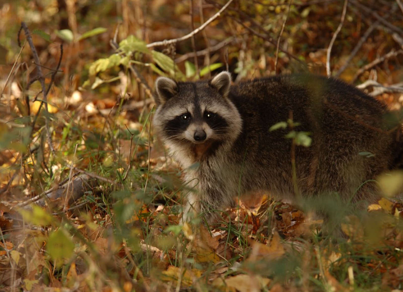
Wild animals, just like humans, may become ill when they contract certain diseases. While some of these diseases are transferable to humans, others are not. Most diseases that human’s contract from wild animals are curable if accurately diagnosed and given appropriate medical attention. If medical attention is not received, some diseases may cause permanent damage to the body or even death.
Rabies
One of the most common diseases found in wild animals is Rabies. Rabies is classified as zoonoses or zoonotic. “Zoonoses” is a term originally used to describe diseases that humans may acquire from domestic animals. This has been modified to include all human diseases that are acquired from or transmitted to any other vertebrate animals, which includes humans.
This fatal disease is caused by the rhabdovirus which in the family of negative-sense RNA viruses. Rabies is also known as an enzootic disease, one that affects or is peculiar to animals of a specific geographic area and can lead to Epizootic outbreaks. Epizootic outbreaks occur when large numbers of wild animals become infected at the same time within a region. Historically, there have been numerous Epizootic outbreaks of rabies in the Southeast, including Alabama.
There are currently four rabies variants in the United States. A variant is a version of something that differs in some way from other forms of the same thing. Rabies variants are found in the skunk, fox, raccoon, and bat. In the late 1980’s to the mid-1990’s, a strain of rabies, common to dogs and coyotes in Mexico, started showing up in Texas. However, wildlife experts conducted a series of oral vaccination bait drops, thus eliminating this variant from the United States.
The two types of rabies virus found in the Southeast are the raccoon and bat variant. These two variants are enzootic with epizootic outbreaks. The general difference between each variant is the carrier. Each variant can lead to death if an infection occurs in other mammalian species. For example, if a raccoon is infected with the bat variant of rabies, or vice versa, a full infection will occur with the infected animal succumbing to the symptoms relatively quickly. Each of the stated animals are not carriers of the other variant. When testing for rabies in a region, it’s important to know what variant you are dealing with to determine the proper response.
One mammal that seems to be resilient to the rhabdovirus, and thus, rabies, is the opossum. According to the Field Manual of Wildlife Diseases in the Southeastern United States, by William R. Davidson and Victor F. Nettles, “opossums historically have been exceedingly rare wildlife hosts of the virus. Opossums are highly refractory to infection by rabies virus, and the amount of virus required to experimentally infect opossums is extremely high. This factor may be responsible for the virtual absence of rabies in this species.” Although the virus is rare in opossums, if bitten, one should contact local health departments.
How can you tell when an animal has an active rabies infection? According to Davidson and Nettles, “Raccoons and other animals with rabies typically have marked changes from normal behavior. Often these are classified into two forms: dumb rabies and furious rabies. Dumb rabies, characterized by aimless wandering, lethargy, incoordination, weakness in hind legs, paralysis, and loss of awareness, is the most common form in raccoons.” (p 139) Furious rabies may cause the animal to lash out in unprovoked attacks on any moving object and may also lead to self-mutilation. In either case, the end is characterized by convulsions, coma, and death.
Although these physical symptoms may constitute suspicion of an active rabies infection, final diagnosis and confirmation is only achieved through testing brain or brainstem tissue. Quarantine and observation periods are used with domesticated animals, but this method is unreliable with wild animals because of the virus’ incubation period. Suspected wild animals should be euthanized, double bagged using rubber gloves, and sent to the health department for testing. It is important that the cranium or brain not be destroyed. Contact the health department before submitting a specimen to make sure the appropriate samples and all pertinent data is included to conduct reliable testing.
Many well-intentioned individuals have also provided a means for the raccoon variant of rabies to become established in new areas. When humans reintroduce exterminated wildlife to historical home ranges, the population increases and presents a new genetic pool to the current population. Additionally, improper isolation and quarantine of an animal for the purposes of diagnostic pathogen testing can wreak havoc on unsuspecting wildlife populations. Even though infected raccoons have been known to swim large river systems and even use bridges to cross major waterways, the main source of translocation of the rabies virus seems to be through human intervention. Relocating raccoons is not recommended as isolated populations of raccoons that do not have confirmed rabies cases can also be subject to possible infection.
Despite the fact that it is illegal, relocating a carrier animal just a few miles from low density areas to more habitat suitable high-density sites could cause an epizootic outbreak. The best solution is not to relocate any raccoon regardless of the situation. Despite the illegality of relocating raccoons across major waterways and county lines, well-intentioned humans and those hoping to increase raccoon hunting success will only aid in the westward migration of the raccoon rabies variant and increase the risk of human exposure to the virus.
The bat variant of rabies is also prevalent in the Southeast region of the United States. Although exposure to and transmission from animal to human is also possible, it is unlikely. Only about 1-3% of all bats have rabies infections. Although less common than the raccoon variant transmission, there have been cases of the bat variant of rabies infecting humans through possible airborne contamination. But keep in mind, this is extremely rare. According to a Center for Disease Control document, two individuals who died from the bat variant had previously been in caves where millions of bats were congregated. Airborne contamination seemed to be the cause of the virus transmission. Having a bat flying overhead at night in your backyard is not considered exposure and is not grounds for preventative treatment.
The best way to minimize exposure to and possible contraction of the rabies virus is to be very careful around any animal, domestic or wild. The most common way rabies is transmitted is through the bite or scratch from an infected animal. As the virus is transmitted through the saliva of the infected animal, contact of the animal’s saliva with open wounds or mucous membranes on a victim constitutes rabies exposure. Scratches from infected animals are grounds for possible exposure as these appendages could have saliva on them that contains the virus. Rabies is not transmitted through blood, feces, or urine from the infected animal. Additionally, according to an article titled, “Twelve Common Questions About Human Rabies and Its Prevention” published in Infectious Diseases in Clinical Practice (2000) 9:202-207, infectious material which has been dried “can be considered noninfectious because the rabies virus is inactivated by desiccation and ultraviolet radiation.” Also, the article states, “rabies cannot be transmitted from an animal that does not have active infection.”
Pseudorabies
Pseudorabies is caused by a virus found in swine called the Suid herpesvirus. This DNA virus, though, has no relation to the rabies virus. Even though domestic and feral swine are the hosts for this disease, it can also infect cattle, goats, dogs, cats, horses, bears, rodents, raccoons, skunks, and many other mammalian species. There are no documented cases of Pseudorabies in humans.
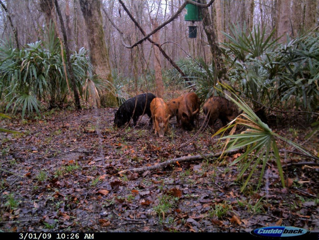
This virus was given the name pseudorabies because the symptoms that non-swine mammals exhibit are similar to the symptoms of those infected with rabies. The virus attacks the central nervous system and animals with an acute infection may have loss of appetite, vomiting, tremors, foaming, blindness, spasms, biting, self-mutilation, and intensive scratching. This virus is also referred to as the “mad itch” disease. Once an infected animal begins showing clinical symptoms, mortality is near 100%.
Swine are the only host to pseudorabies and some swine may survive the infection and be latent carriers. These animals show no symptoms of the virus but can continue to transmit it through many routes. Swine disease transmission is mainly through venereal route but can be transmitted via oral, nasal, digestive or reproductive mucosa. Other transmission can be through nasal and oral contact, contaminated food, water, or structures. Non-swine species transmission is mainly by the latter. Also, feeding on infected carcasses can also transmit the disease and possibly spread it.
Pathological testing is required for pseudorabies confirmation. Florescent antibody testing on brain or tonsil tissue or testing for antibodies in serum can positively determine the presence of the pseudorabies virus. There is no treatment available for pseudorabies and infected non-swine species are considered dead-end hosts because of the high mortality rate.
Swine Brucellosis
Swine brucellosis is an infectious disease caused by the bacterium Brucella suis. This bacterium is found in the reproductive tract and infections can cause spontaneous abortions, low milk production, infertility in sows and affect the testicles in boars. In boar’s, brucellosis can affect their reproductive fertility. Serology testing in feral swine populations around the country has shown this disease is quite common. The main transmission of this bacterium is through reproduction. When this zoonotic disease infects a human, it can be fatal. This bacterium can live for hours outside the host which makes humans more vulnerable for transmission.
Symptoms of brucellosis in humans are very similar to the influenza. Fever, body soreness, headaches, sweating, and fatigue are just a few. An infected person can also have chest and abdominal pains, difficulty breathing, loss of appetite and irritability. If one goes to a physician with any of these symptoms after field dressing a feral hog, they should tell the physician they may have been exposed to swine brucellosis. Often physicians do not test for brucellosis as this is a very uncommon diagnosis, unless they are made aware of the likeliness of the exposure.
When field dressing feral swine, it is highly recommended to take several precautions to avoid this bacterium. One needs to use disposable gloves and a rain suit that can be removed and washed when done. Also, rubber boots are recommended. Be careful when washing out the carcass. Minimize touching the reproductive tracts in both boar and sows when possible. When completed, wash the area in hot soapy water if possible or spray area with a 10% bleach mixture.
The Alabama Cooperative Extension Service posted two feral swine field dressing tutorial videos on their You Tube page and are quite informative for anyone planning to field dress a feral hog. One video is a traditional method of hanging a hog and the other is the “quick tailgate method.” Both videos demonstrate proper personal protection equipment and how to mix a salted ice solution for quickly lowering the temperature of the meat so it will not spoil. These videos discuss swine brucellosis and preventative measures that should be taken when field dressing any feral swine.
Cutaneous Fibroma
Cutaneous Fibroma is a simple wart caused by the papilloma virus, but it can look hideous to people who have never seen it on a white-tailed deer walking through the woods. Cutaneous fibromas are often referred to as deer warts. They can be as large as golf balls or small as a grape. These tumors are black to brownish with a rough texture. The warts are attached only to the skin, so they move freely from the muscle. If the fibromas don’t make it difficult to breath or eat, the deer will have a healthy life.
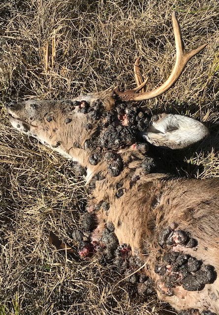
Deer contract this virus through numerous pathways. Biting insects can spread the virus from deer to deer. Sparring bucks can also transmit the virus. A deer with an open wound, walking down a trail and rubbing against a stick that contains the virus, could cause this temporary inconvenience. Studies suggest that after a short inoculation period the warts may disappear in a few months. Deer that have been exposed and recovered can develop an immunity to the virus.
The two main concerns when one sees a deer with warts is whether it is suitable for eating and its impact on the herd. Deer with this virus are edible and palatable. The virus does not affect the meat in any way so it can be processed for human consumption like any other deer. Once the skin is removed the carcass looks like that of any other harvested deer. This virus does not transmit to humans so field dressing is safe. It is recommended though, to always use rubber gloves when field dressing any animal.
Removing, or culling, a deer with warts to keep from spreading the virus will have little to no impact on the herd. As stated earlier, the deer’s immune system will take care of the virus and in a few months they will go away. The virus is already in the environment and that young buck with warts may grow to be a trophy in a few years. There may be many deer with cutaneous fibromas walking around in the woods but are just never identified.
Canine Distemper
Canine Distemper is a disease in the paramyxovirus family which also includes measles, mumps, newcastle disease, parainfluenza, and RSV (respiratory syncytial virus) which is a major cause of bronchiolitis and pneumonia in infants and children. Raccoons, gray foxes, coyotes, and unvaccinated domestic dogs can get canine distemper. Canine Distemper disease has also been confirmed in skunks and minks as well.
This respiratory virus is commonly transmitted from animal to animal through secretions such as nasal and eye discharge. Transmission can also occur from contact with airborne droplets. Contact with the feces or urine of an infected animal can also transmit the disease. Symptoms of infected animals include coughing, sneezing, convulsions, chewing, and thickening of the foot pads and nose. Animals that have this virus are usually emaciated and have diarrhea.
Canine distemper has a nearly 50% mortality rate among raccoons and coyotes. The mortality rate for mustelids is near 100%. Animals that recover can still spread the virus for weeks. The virus is not stable in the sun or heat, but in colder environments or shade it can be viable for a long period of time and even up to weeks in freezing temperatures.
In epizootic outbreaks, this virus can drastically reduce the local populations of raccoons. Infected animals can have the same mannerisms of a rabid raccoon and are often misdiagnosed as rabid. Positive diagnosis to confirm canine distemper can be done by serology. If a field necropsy is done, specimens of the brain, lung, liver, and other organs can be used for the fluorescent antibody testing used to confirm Canine Distemper.
This disease cannot spread to humans but can be profoundly serious to unvaccinated pets. Most domestic dogs get their vaccine as puppies which is important as there is no treatment for distemper. Those animals that survive the infection may have medical issues their entire life.
Conclusion
The diseases detailed in this article make up just a small fraction of wildlife diseases that are present in the world around us. Some have no effect on humans or domestic pets while others can be fatal if not properly treated. Some diseases have no treatment at all, and supportive care is all that can be done to unvaccinated animals. Epizootic outbreaks can have a drastic effect on local populations of certain wildlife species, but usually do not have long-term consequences. It is not logistically possible to attempt to control most wildlife diseases. Therefore, epizootic outbreaks will occasionally happen. Depending on how significant the mortality may have been, reproduction of the surviving animals, along with immigration, usually repopulates an area within a year. Wildlife diseases, just like human diseases, are a part of our existence. Our best strategy for dealing with them is to educate ourselves and engage in practices and behaviors that minimize the risk of infections.