

In Part 1 of this series, we provided an overview of several bacterial, viral, and chemical wildlife diseases. In Part 2, we will focus on some important parasitic and fungal agents, with regards to game and non-game wildlife species. In addition we will examine an important disease of wild deer that does not fit into any of the typical causative categories. At the conclusion of this series we will have provided an overview of some of the most important modern diseases in US wildlife.
Wildlife Parasites
Parasitism is defined as “an interaction of species populations in which one organism (the parasite) lives in or on another organism (the host)”. A parasite obtains something it needs (food, shelter, or water) from its host. Technically viruses and bacteria could be referred to as parasites, but for the purposes of this discussion we will only include multicellular organisms, as we have already covered bacterial and viral agents in Part 1. Virtually every living complex organism has parasites, and thus the list of parasites in North American wildlife is obviously very extensive. For example, deer alone have been shown to harbor over 130 species of parasites. We will therefore discuss some of those that are associated with the most popular game and non-game animals.
Parasites can be ecto-parasitic (living externally on the host) or endo-parasitic (living within the host). Ecto-parasites are generally less apt to cause direct harm to the host, although they can serve as vectors for more serious diseases. They are usually after only a small portion of the host animal’s blood, but sometimes they can collectively steal enough of the host’s energy to cause death, in even large animals. Ticks, for example, are a well-known ecto-parasite that can transmit several serious zoonotic diseases like Lyme and Rocky Mountain spotted fever. In humans both of these bacterial diseases are quite serious, but they typically do not pose much of a health risk for most wildlife hosts. Ticks can have a direct impact on even large herbivores however if the infestation is severe enough. Individual moose and deer have been found to sometimes harbor winter loads of over 25,000 ticks. This many blood-sucking parasites can cause emaciation and starvation, especially in colder regions where the harsh winter months can make food scarce. Other ecto-parasites like fleas, mites, and mosquitoes can also serve as vectors for bacterial and viral diseases in wild populations but for the most part they are just an annoying part of life. And although extreme ecto-parasite infestations can pose a threat, the more significant parasites are generally endo-parasites.
One of the largest groups of endo-parasites is the roundworms (nematodes). Almost every animal species is vulnerable to some species of parasitic nematode. Nematodes are long, un-segmented worms and since many species are relatively large, they are sometimes seen in infected game species by hunters during the cleaning process. After an infected game animal is shot and its biological functions cease, roundworms can sense the change and may randomly migrate out of normally infected sites and into the more visible major body cavities. Typically nematode parasites do not kill their normal hosts (they are more likely to cause more harm in abnormal hosts) but they can cause certain biological stresses. The large deer lungworm for example lives in the lungs of its host but generally causes no negative symptoms at low levels. More severe lungworm infestations (often associated with high density deer populations) can lead to weakness and respiratory problems however. Intestinal roundworms are another example of common nematode parasites, and affect many wildlife species. Gastro-intestinal nematodes (like the large stomach worm) in deer infect the abomasum and intestinal tract and can reach levels that cause reduced growth rates, poor antler development, reduced reproductive potential, and lowered immunity. These intestinal roundworms (generally harmless to humans) have a relatively simple life cycle in ruminants like deer. In the intestinal tract, the adults produce eggs which are deposited back onto foraging areas with the scat of the deer. The eggs develop into larvae that crawl onto vegetation or into water sources where they can be ingested by new hosts or re-ingested by currently infected deer. These repeated re-infestations can lead to heavy loads in local deer populations, especially in densely populated areas. These parasites typically are driven by natural cycles and usually don’t significantly limit wild populations, however there is actually a fairly new deworming product on the market for treating local deer herds in some situations. Fenbendaxole (SafeGuard) has been used in domestic animals for several years. It has recently been approved by the FDA for use in a pelleted form for wild deer, with no adverse reaction to non-target animals such as turkeys or wild rodents (Bliss, 2015). The pellets are spread onto foodplots or other feeding areas (at a recommended target dose rate of 3.4 mg/lb) over a period of a few days, usually used in conjunction with camera monitoring to help determine whether dosage is obtained. Wild deer deworming is not a critical need in normal circumstances and treatment wouldn’t be advised unless recommended by a local deer biologist.

Another common example of a nematode is the canine heartworm (Dirofilaria immitis). This is the same species that infects domestic dogs, but it can also affect foxes, coyotes, wolves, raccoons, muskrats, and cats. Heartworms are spread from host to host by the bite of a mosquito. Although this slender thread-like parasite is called a heartworm, they will eventually also spread into the pulmonary arteries and lungs of the host and ultimately can cause congestive heart failure. In domestic dogs, prevention is the best control and the current approved treatment for existing infections is a drug called melasamine dihydrochloride. However in wild animal populations there is no current treatment protocol for heartworm. Fortunately occurrences are usually not overly common in free-ranging mammal populations.
Just as in mammals, birds are also highly susceptible to roundworm parasites. As an example, one study found 34 species of nematodes in 27 species of waterfowl (McDonald, 1974). About 97% of all the sampled birds (N=415) were infected at some level. One particular specimen (a whistling swan) harbored eight separate species of nematode parasites. The proventricular worm, aka stomach worm, (Dispharynx nasuta) is one of the most common species seen in songbirds and gallinaceous birds in the United States. Crows, bluejays, gray catbirds, American robins, wood thrushes, and many more species of passerines, along with virtually all upland game bird species (including woodcock, ruffed grouse, and quail) are susceptible to proventricular worms. Typically juvenile game birds have higher intensities of infection as compared to adults. Wild turkeys for example usually become infected within the first week of life in areas that are infested with this nematode. As with many other gastro-intestinal nematodes, adult proventricular worms produce tiny tough-shelled eggs that pass out of the final host with their droppings. However, instead of waiting on vegetation the tiny larvae typically use an intermediate host; the pill-bug (rolly polly). Young turkey poults readily ingest the infected pill-bugs as they are encountered and the cycle starts over again. In turkeys and most other birds the symptoms can be mild to severe, depending on the intensity of infection. This species can cause emaciation and a weakened state, making individuals more at risk of being caught by predators. In severe cases (dozens of adult worms) it can cause swelling and blockage of the proventriculus and lead to starvation. This parasite has been determined to be the most important limiting factor in ruffed grouse populations in certain local regions (Michigan DNR, 2015).

Cestodes (tapeworms) are another endo-parasite one will probably encounter from time to time while dressing wild game or examining wildlife. Tapeworms are a completely different type of organism from nematodes. They are flattened and segmented in appearance with a “head” equipped with tiny hooks for affixing themselves in the primary host’s digestive system. Although there are many, the most commonly observed species of tapeworm are probably within the Taenia and Echinococcus genera and have a two-host life cycle. The adult tapeworms live in the gut of a carnivore like a dog, coyote, bobcat, or mountain lion. These adults produce eggs which pass out of the animal and contaminate vegetation and herbaceous plants. The intermediate herbivore host (deer, elk, moose, rabbit, etc.) ingests the eggs and upon hatching in the stomach, the tapeworm larvae begin to travel through the bloodstream. The larvae will wind up in the muscle tissue of the herbivore where they will encyst and wait. The cycle is completed when the infected herbivore (along with the encysted larvae) is eaten by one of the aforementioned predators. Deer and other intermediate hosts are not usually weakened by tapeworms, although heavy infection in canids or other predators can sometimes cause stress. Humans are not a normal host for these species of wildlife tapeworms but can become infected occasionally from exposure to eggs from a contaminated dog or other primary host (not deer). Cooking destroys the cysts but one should also be careful about feeding pets meat scraps when dressing or butchering herbivores. They can easily become infected if there are tapeworm cysts in the meat.
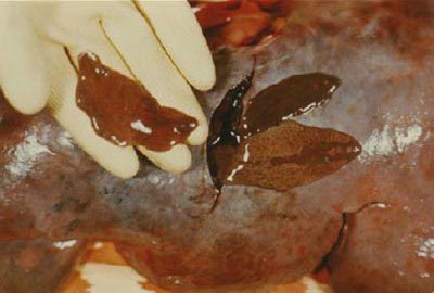
Other major endo-parasites of white-tailed deer worth mentioning here are the deer liver fluke, brainworm, and nasal bots. Another formerly important species called the screw worm fly was a natural parasite until its eradication from the US in the early 1980’s. The liver fluke (especially Fascioloides magna) is a flatworm and looks like a big flattened leech up to about 4″ long. The normal life cycle involves two hosts; deer (primary) and snails (intermediate). In the normal deer host the fluke rarely causes sickness or major population issues, although individuals could experience liver damage under heavy infestations. Occasionally however domestic livestock will become infected and the fluke can easily kill sheep. Cattle are a little more tolerant of the parasite. Fluke-infected venison is reported to be safe for human consumption and no direct human threats are presented. Although it is a nematode, the brainworm (or meningeal worm) has a similar life cycle, involving white-tailed deer and snails or slugs. This parasite does cause some damage to the central nervous system in deer but again does not typically kill the normal host. In areas where the ranges of moose and white-tails have recently merged however (due to expanding deer populations), the brain worm is becoming a significant concern. In moose, the damage is much more severe and death is common post-infection. Declines in southern moose populations are predicted over the next several years in these overlapping areas (Murray et. al., 2006). Nasal bot (Cephenemyia spp.) larvae are essentially large maggots that infect a large percentage of North American white-tail deer and to a lesser degree, mule deer and elk. Interestingly these parasites seem to be absent or very rare in a geographical band that runs through the Midwest down into Tennessee and Alabama. They live in the nasal passages, sinuses, and deep pouches in the back of the host’s mouth. These parasites are only a minor nuisance to healthy deer, even when the infestation is heavy. The bots fall out when they near maturity and then pupate in the soil. The adult (fly) then emerges and, after mating, will seek out a deer to complete its life cycle. In flight the female bot fly deposits her tiny new larvae (the eggs hatch inside the fly) in the moist warm nasal canal of an unsuspecting deer. Again with this parasite, no concerns exist regarding human consumption of infected deer meat. A very similar species of bot fly commonly infects squirrels (and occasionally other rodents and rabbits) across the country. However “warble fly” larvae create and maintain a breathing hole and live under the skin of the squirrel, instead of in the nasal passages. There the larvae can irritate the local area and causes discomfort in the host, but again it is generally not life threatening. The larvae usually drop out in the fall to pupate in the same manner as the nasal bot, and the host quickly heals.
Fungal Diseases in Wildlife
The final major category of disease-causing organisms involves fungi. For the past fifty years or so, fungal wildlife diseases have been on the rise, due in part to an increased global trade and travel industry. In fact, modern fungal diseases are thought to be the most significant threat to wildlife diversity across the world. Three notable “new” fungal diseases that have become concerning within the last couple decades are White-nose syndrome in North American bats, Batrachochytrium dendrobatidis in amphibian populations across much of the world, and snake fungal disease. White-nose syndrome or WNS (caused by the cold-loving cave fungus, Pseudogymnoascus destructans) has spread from New York to 25 known states and five Canadian provinces since 2006. As we discussed in a previous Wildlife Trends article, the WNS fungus colonizes and spreads on hibernating bats and typically causes mass mortality events due to constant awakening of the bats in their hibernacula. The disease has caused severe declines in several species of cave-hibernating bat species in the past nine years and is expected to completely shift the entire ecology of bats in the United States.
Batrachochytrium dendrobatidi (or Bd) has been causing similar alarming declines in frogs and other amphibians in Asia, Central and South America, and several other parts of the world. Unfortunately it is probably only a matter of time before the southern US experiences significant amphibian die-offs due to this fungus, which attacks the moist outer skin of its victims.
Snake fungal disease is a brand new concern and research scientists are still trying to figure it out. Since about 2006, it has been reported from nine eastern and Midwestern states (including Tennessee and Florida in the South), but it is likely to be more widespread. At the moment it has only been found to affect snakes species and it causes skin separation, subcutaneous nodules, and other issues (USGS, 2015). It appears that at least one fungal organism (Ophidiomyces ophoidiicola) is to blame in most cases but there are often several fungal species isolated from diseased individuals. It is not clear as to what extent it may affect populations but we can expect to hear more about this strange fungal disease as more is learned in the coming years.

A better known fungal disease that can affect humans and several wildlife species is aspergillosis (caused by Aspergillis spp. fungus). It is serious but quite rare in humans and usually occurs in the lungs of immunocompromised individuals. In wild animals, it is most important in birds (especially waterfowl). They typically become infected with the fungus by feeding on old moldy grains or other fungus-contaminated food sources. Numerous die-offs due to aspergillosis have been reported in ducks over the past several decades. For example 500 mallards were killed by this disease in Iowa in 2005, and in 2006 over 2,000 were found dead in Idaho. Both events were due to feeding on moldy waste grain and corn. This emphasizes the importance of responsible supplemental feeding. If you feed corn or other dried grains to wildlife, make sure you maintain clean quality feed and feeders. In fact, this tip applies to reducing the risk for many of the other types of diseases we have discussed in this series. Any time disease agents and high concentrations of wildlife occur (such as in supplemental feeding areas), there is increased risk for disease transmission.

Other Diseases
There are some diseases that do not fit nicely into any of the above categories. One such example is Chronic Wasting Disease (CWD) which is known to affect deer, elk, and moose. CWD was detected in wild mule deer and elk in parts of Colorado and Wyoming in the mid-1980’s. Since then it has spread to other adjacent western states like Nebraska (2001), Utah, South Dakota, and Kansas (2006), and into Wisconsin, Illinois, and New Mexico (2002). In 2005, it was found in wild white-tailed deer in New York and West Virginia, Michigan in 2008, and most recently (2011) in Maryland. This strange disease is a form of transmissible spongiform encephalopathy (similar to “Mad Cow Disease” in cattle and “Scrapie” in sheep) and it is believed to be caused by an abnormal form of a protein called a “prion”. My graduate professor described prions as “folding proteins” or “helper proteins”. In their normal healthy state, they most often function in the moving of molecules across individual cell membranes. Prions are not living organisms (they do not even contain DNA or RNA like viruses), but upon contact, the abnormally folded form rapidly converts normal proteins into the disease-associated misfolded formation. Newly converted proteins can then, in turn, infect more normal proteins, initiating an exponential replication of abnormal prion molecules. The resulting structures accumulate in infected neural tissues and cause widespread death of cells and a sponge-like appearance at the affected site (initially in the brain and spinal cord). As the disease progresses during the incubation (wasting) period, the abnormal prions can spread via the peripheral nervous system into the muscle (meat) of the animal. It is inconclusive at this point whether CWD can actually infect humans, however the CDCP strongly advises against consuming some deer and elk tissues (brain, eyes, spleen, etc.) from areas where the disease has been identified.
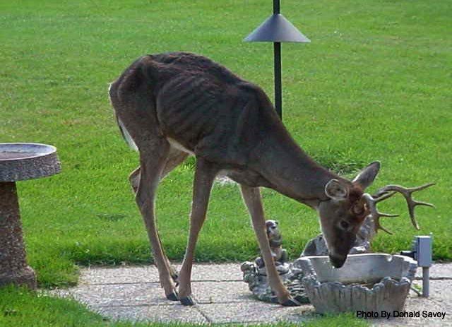
Prion proteins are extremely stable, and therefore are very resistant to chemical and physical denaturation. In other words, they can persist in the soil or grass for long periods of time and make disposal and containment of infected animals very difficult. High temperatures (associated with cooking) typically do not affect prions either. This is another reason not to take a chance on eating any questionable meat from areas with high CWD contamination. In humans, there are four known prion-caused diseases (Creutzfeldt-Jakob Disease, Gerstmann-Strausster-Scheinker Syndrome, Fatal Familial Insomnia, and Kuru) with physiological impacts similar to CWD, but that are not caused by the CWD prions. The first three of these are either inherited (familial) or acquired through mutation or human-to-human tissue transplants, while Kuru is associated with a ritual called “mortuary cannibalism” in the tribal regions of Papua New Guinea. This strange disease was perpetuated by the custom of eating human corpses (especially portions of the brain) and it even reached epidemic proportions in the mid-1900’s!
In summary, wildlife pathogens and diseases are complex and diverse. Obviously we could not cover all of the pertinent diseases wildlife managers may encounter in our native species, but with the conclusion of part 2 of this series we have provided a thorough overview of some of the most important modern game and non-game disease agents. Just as in humans, diseases in wildlife are dynamic and can change over time. However by understanding stressors, pathogenic organisms, and potential control measures, we as wildlife managers can make better decisions regarding the wildlife populations for which we are responsible.
References
Bliss, D. H. The control of gastro-intestinal nematode parasites of hooved wildlife in North America. Mid America Ag research technical Bulletin. Retrieved online March 25, 2015.
McDonald, M. E. 1974. Nematode parasites of waterfowl from western United States. USDI Fish and Wildlife Service Publication (Wildlife Disease), Issue 64.
Michigan Department of Natural Resources. Michigan Wildlife Disease Manual (Deer Nose Bots). Retrieved online April 1, 2015.
Michigan Department of Natural Resources. Michigan Wildlife Disease Manual (Proventricular or Stomach Worm). Retrieved online March 25, 2015.
Murray, D. L., Cox, E. W., Ballard, W. B., Whitlaw, H. A., Lenarz M. S., Custer, T. W., Barnett, T., and Fuller, T. K. Pathogens, nutritional deficiency, and climate influences on a declining moose population. Wildlife Monograms 166(1):1-30
US Geological Survey. National Wildlife Health Center. Snake Fungal Disease. Retrieved online April 2, 2015.